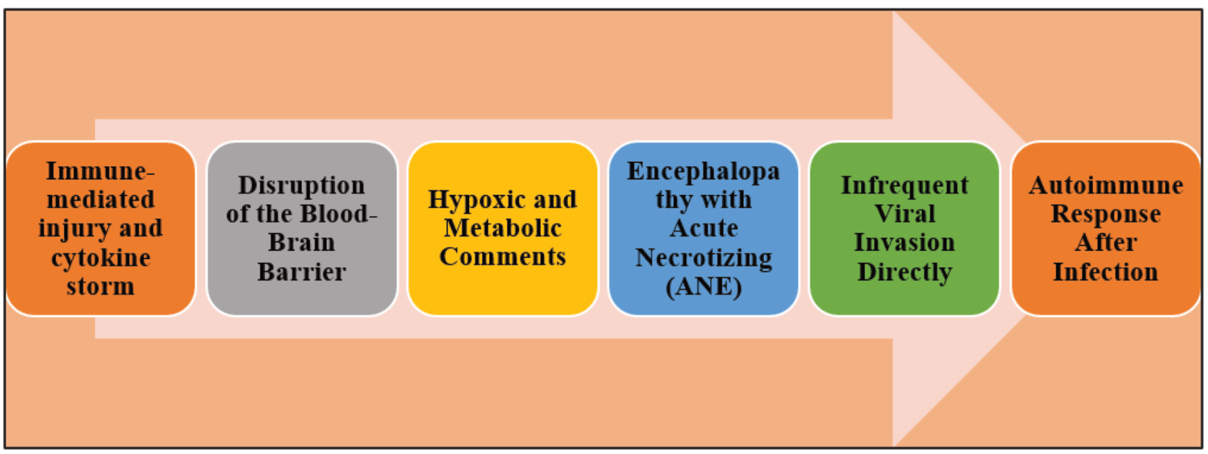
The manifestation of encephalopathy associated with H3N2 influenza virus infection underscores the growing concern about the virus's neurotropic potential, especially in vulnerable populations such as children and the elderly. While influenza is predominantly known for its respiratory symptoms, increasing clinical evidence reveals that it can cause severe Central Nervous System (CNS) complications ranging from mild confusion to Acute Necrotizing Encephalopathy (ANE). The pathophysiology of such encephalopathies appears to be multifactorial, involving immune-mediated mechanisms, cytokine storms, metabolic disturbances, and, rarely, direct viral invasion of the CNS. Elevated inflammatory cytokines such as IL-6 and TNF-α disrupt the Blood-Brain Barrier (BBB), permitting the infiltration of immune mediators and toxins into the brain parenchyma. This often leads to neuroinflammation, cerebral edema, and in severe cases, symmetrical brain lesions particularly in the thalami and brainstem. Case studies discussed in the review highlight the heterogeneity of clinical presentations and outcomes, ranging from full recovery to fatal cerebral failure. Most notably, paediatric cases with ANE showed rapid deterioration with elevated intracranial pressure and multi-organ dysfunction. Conversely, elderly patients displayed post-infectious autoimmune encephalitis, with partial or complete recovery upon immunosuppressive therapy. These cases affirm that H3N2-induced encephalopathy is not only a complication of infection but a consequence of an aberrant host immune response. Despite these insights, the rarity of viral detection in Cerebrospinal Fluid (CSF) challenges direct viral attribution and necessitates further research into molecular mimicry and immunogenetic susceptibilities. Timely diagnosis through MRI, CSF analysis, and early supportive care with antivirals and corticosteroids remains critical. Greater awareness, rapid intervention, and targeted management strategies are imperative to minimize the neurological morbidity and mortality associated with H3N2 influenza (Tsai and Baker, 2013).
Earlier studies explored the risk of Encephalopathy associated with influenza virus. In a case, in the emergency room of Hainan General Hospital, an earlier fit 3-year-3-month-old female infant weighing 12 kg who had not received the influenza vaccine presented with a past of hoarseness for 3 days, pyresis, diarrhea for half a day, and three events of seizures. On Day 1, the infant started coughing with sounding hoarse, so oral antibiotics and cough suppressants were administered. On Day 4, she was initially brought into the outpatient clinic with pyresis, puking, and diarrhea. However, due to the onset of a high body temperature and three generalized seizures, the infant was sent to the Pediatric Intensive Care Unit (PICU) the following day after getting emergency care with diazepam, phenobarbital, cefmetazole, and methylprednisolone. Each generalized convulsion lasted about 3 min and had symptoms like loss of consciousness, upward gaze, and rigidity of the extremities. The kid dozed off in between seizure bouts. Medication could be used to control each convulsive episode. Her Blood Pressure (BP) was 92/62 mmHg, respiratory rate was 24 breaths per minute, body temperature was 36.2°C, and heart rate was 132 beats per minute. Midazolam was used to sedate the infant continuously. Her respiration was regular, respiratory sounds were coarse in both lungs, and sputum noises could be heard while she was under anesthesia. Her bilateral pupils were equal in size and circular with blunted light reflexes. Meningeal irritation was also negative. None of the additional investigations revealed any anomalies.
A nasopharyngeal exudate tested positive for H3N2 after the patient was admitted to the PICU. Regular blood tests revealed low levels of serum potassium, elevated levels of blood lactate, alanine aminotransferase, and aspartate aminotransferase, normal levels of blood ammonia and copper cyanide, and signs of coagulation failure. pH was 7.34, PaCO2 was 29.7 mmHg, PaO2 was 98.5 mmHg, HCO3 was 16 mmol/L, and base excess was 7.8 mmol/L according to blood gas analysis. The Cerebrospinal Fluid (CSF) was also colourless and transparent, with a normal glucose, chloride, and cell count, as well as a noticeably high protein concentration (1.69 g/L; reference range, 0.15-0.45 g/L). Results from tests on the cellular and humoral immune systems as well as the findings from a cerebral computed tomography examination were all normal. The child was identified as having severe influenza A and encephalopathy caused by the influenza virus. She received mannitol for dehydration to lower her intracranial pressure, midazolam for continuous sedation, an ice pack for continuous physical cooling, ceftriaxone (100 mg/kg per day for infection control), dexamethasone for anti-inflammation therapy, correction of hypoglycemia, prothrombin complex infusion for improved coagulation, and liver protection therapy. A child's echocardiogram was normal around 5 hr later, but a high cardiac troponin T concentration (0.257 g/L) was found, which may have indicated myocardial injury. The infant was then given calcium dibutyrylladenosine cyclophosphate and Vitamin C via an intravenous drip. She showed a poor response to painful stimuli and shallow, erratic respiration around 9 hr later. Glasgow coma scale score for the youngster was 4, with the best eye response (+1), best verbal response (+1), and best motor response (+2). At 39.0°C, central respiratory failure was thought to be present. Following invasive ventilation and endotracheal intubation, the kid received symptomatic care in order to keep the internal environment stable. Dynamically tracking the child's internal environment during the therapy period revealed severe hepatic impairment, coagulation malfunction, myocardial impairment, acidosis, and hypokalemia in the biochemical blood test findings. She was given low-dose dopamine (4 g/kg/min) after showing signs of deteriorating BP (80/60 mmHg), oliguria, and hyperlactatemia 14 hr later. A little over 33 hr later, the kid manifested signs of brain failure, including unstable blood pressure, bilaterally dilated and fixed pupils, loss of the light reaction, and a progressive rise in blood glucose levels. The child passed away 48 hr after being admitted (Li et al., 2023).
On Day 1, a 24-kg, 8-year-old child who had not received the influenza vaccine developed a fever. She had a 41.3°C body temperature peak. Along with the fever, other symptoms were a headache, sore throat, runny nose, and dizziness. Cefixime, an oral solution of aminoglutethimide, and acetaminophen were used to treat her. She continued to have frequent high fevers, and on one occasion even went into a convulsion, thus no recovery was seen. On Day 3 following therapy, she went to Hainan General Hospital and was given intramuscular injections of phenobarbital, methylprednisolone, and an antipyretic medication. Her temperature was 41.3°C, respiration rate was 38/min, heart rate was 181 beats per minute, and blood pressure was 83/63 mmHg when she was examined. The patient appeared sleepy, and her entire body was covered in skin mottling. Her breathing was regular, she had round, equal-sized pupils on both sides, a blunted light reflex, coarse respiratory sounds in both lungs, audible phlegm, and dry rales. Meningeal irritation was also negative. Reflexes in the nerves and other tests revealed no abnormalities. Testing of a nasopharyngeal exudate samples after admission to the PICU revealed H3N2 positivity. The white blood cell count was 6.31 109/L, with 70.6% neutrophils, 27.9% lymphocytes, and 1.3% monocytes, according to routine blood testing. At 4.07 mmol/L, the lactic acid concentration was high, but there were no abnormalities in the CSF. The child was identified as having severe influenza A and encephalopathy caused by the influenza virus. She received mannitol for dehydration to lower her intracranial pressure, midazolam for ongoing sedation, an ice pack for ongoing physical cooling, oseltamivir for ongoing antiviral treatment (60 mg nasogastrically twice daily), ceftriaxone for ongoing anti-infection therapy (100 mg/kg per day), dexamethasone for ongoing anti-inflammatory therapy, and fluid to maintain the internal environment. Prothrombin time was 25.5 sec, activated partial thromboplastin time was 63.9 sec, fibrinogen concentration was 1.74 g/L, and the international normalized ratio was 2.67, according to coagulation testing. Alanine aminotransferase was detected by liver function tests at 2440.6 U/L. The child's echocardiogram was unremarkable 20 hr later. A second echocardiographic examination performed 37 hr later, however, revealed that the left ventricular wall motion had decreased. The child's heart rate was 151 beats per minute, his blood pressure was 64/48 mmHg, and his capillary refill time was 5 s. Dopamine (8 g/kg/min) and adrenaline (0.2 g/kg/ min) were continuously infused into the patient. Right cerebral hemisphere circulation was terminated, left cerebral hemisphere circulation was still present, and posterior circulation was also terminated, according to cerebral vascular Doppler ultrasound. Electroencephalography revealed evidence of brain death 48 hr after the patient's death, and cerebrovascular Doppler ultrasound demonstrated complete cessation of all cerebral circulation (Dou and Li, 2022).
A, 70-year-old male patient who had previously experienced sinusitis and nasal polyps arrived at the emergency room with a headache that had been present for two days but no prodromal respiratory symptoms in the previous two weeks. He had never received an influenza vaccination before. The patient arrived at the emergency room with a strong, persistent headache but no vomiting, no loss of consciousness, or any convulsions of the limbs. Clear consciousness, fluent speech, equal pupils, a pupil light reflex that is positive, full eye movement, the absence of diplopia or nystagmus, bilateral facial symmetry, a positive neck stiffness, a negative Kerning sign, a positive Brudzinski sign, and bilateral Babinski signals were all findings of the neurological examination. Arm and leg muscles are strong and under normal strain. Regular blood and biochemical tests, as well as his chest computed tomography, revealed inconsequential results. For suspected infectious meningoencephalitis, intravenous ceftriaxone and ganciclovir were started as broad-spectrum antibiotics. The patient was transported to the Department of Neurology on the third day after being admitted to the emergency room due to a fever that reached 39°C. No unsettling symptoms appeared upon hospitalization. Clear consciousness, fluent speech, equal pupils, a pupil light reflex that is positive, full eye movement, the absence of diplopia or nystagmus, bilateral facial symmetry, a positive neck stiffness, a negative Kerning sign, a positive Brudzinski sign, and bilateral Babinski signals were all findings of the neurological examination. Arm and leg muscles are strong and under normal strain. White blood cell count (8.69 109/L) was within acceptable limits, but aspartate aminotransferase (80 U/L) and alanine aminotransferase (157 U/L) values were abnormally high. A CSF analysis revealed elevated total protein levels of 163 mg/dL, an elevated total cell counts of 720 106/L, an elevated CSF white blood cell count of 650 106/L (42% polymorphonuclear leukocytes, 58% lymphocytes), elevated CSF immunity (IgG=221mg/LIgG=221 mg/L, IgA=42.7mg/LIgA=42.7 mg/L, albumin = 750 mg/L, and IgM=1.16 mg/L), decreased glucose of 2. From metagenomic next-generation sequencing, influenza A H3N2 was found in the CSF. SARS-CoV-2, Herpes simplex, the spectrum of autoimmune diseases, and the spectrum of demyelinating disorders of the central nervous system were not found in the CSF. On the second day of admission to the Department of Neurology, treatment with oral oseltamivir at 75 mg twice daily was started. This was done in light of the MRI findings, clinical presentation, and CSF analysis, which led to the diagnosis of influenza A (H3N2) associated meningoencephalitis. In addition, oral methylprednisolone dosage was gradually tapered off at the same time as intravenous pulse therapy (80 mg per day for 5 days).
The patient started to gradually improve after a 5-day course of oseltamivir and methylprednisolone, and was fully recovered by
day 13. Before leaving the hospital, the CSF underwent a control examination that revealed total protein levels of 52 mg/dL, total cell counts of 40 106/L, CSF white blood cell counts of 30 106/L (13.3% polymorphonuclear leukocytes, 86.7% lymphocytes), CSF immunity (IgG=62.7mg/LIgG=62.7 mg/L, IgA=9.2mg/LIgA=9.2 mg/L, albumin=283 mg/L, IgM=0.0 The outcomes mentioned above are improved over the earlier ones. He was released without experiencing any more neuropsychiatric or encephalitic symptoms. He was given instructions to gradually go off of oral methylprednisolone. The patient experienced a headache and fever again a month later.
When questioned, the patient admitted that he had stopped taking methylprednisolone on his own and had disregarded the doctor's instructions. The right temporal occipital lobe and both choroids clearly showed aberrant enhancement on a contrast-enhanced coronal T1-W imaging. An elevated total protein level of 1,030 mg/dL, a total cell counts of 220 106/L, a white blood cell counts of 190 106/L (32% polymorphonuclear leukocytes, 68% lymphocytes), an elevated CSF immunity level (IgG=113mg/LIgG=113 mg/L, IgA=17.8mg/LIgA=17.8 mg/L, albumin=560 mg/L, IgM=3.27 mg/L), normal glucose at 2. Additionally, there was no microbial development as seen by the negative Gram-staining results. Both the CSF neuronal/inflammatory antibody test and the CSF metagenomic next-generation sequencing yielded negligible results. It was decided to commence intravenous methylprednisolone pulse therapy (500 mg daily for 3 days), followed by a slow tapering of oral methylprednisolone, in order to treat non-specific autoimmune encephalitis brought on by viral infection. By day nine, the subject had fully healed and was released with no signs of relapse. He was instructed to continue the gradual reduction of oral methylprednisolone and to return in three months, if he was pain-free (Li et al., 2023).
A 16-year-old Pune resident who had previously been healthy was hospitalized on August 24th, 2009, to the infectious disease unit with complaints of fever, cough, and rhinorrhea as a case of suspected influenza A (H1N1). She was referred to our tertiary care hospital because she was having trouble breathing. Patient was feverish upon admission to our hospital, but all other vital signs were in the normal range. Her systemic evaluation came out normal. The patient experienced a quick start of altered consciousness on the second day of their stay. She was febrile, had a pulse rate of 116/min, and had a blood pressure reading of 100/70 mm Hg. There was no neck rigidity on the neurologic examination. Despite experiencing severe agony, the patient was not responding to spoken directions. Plantar extensors on both sides had deep tendon responses. There were no symptoms of a focal neurologic impairment. It was usual to look at different systems. The patient afterwards experienced generalized tonic-clonic seizures. The patient needed mechanical ventilation and intubation. A normal urinalysis, chest radiograph, and electrolyte, creatinine, bicarbonate, and glucose levels were all part of the initial laboratory assessment. The platelet count was 62000/mm3, the aspartate aminotransferase and alanine aminotransferase levels were 60IU/L and 85IU/L, respectively, and the white blood cell count was 16,500/mm3 with 80% neutrophils. Time to thrombin was 42/15. According to an MRI, there were bilateral white matter-dominant hyperintensities in the cerebellum and thalamus. Due to indicators of increased intracranial tension, lumbar puncture was postponed. The patient's throat sample tested negative for influenza A H1N1. Herpes simplex, dengue, and Japanese encephalitis research, among others, came up empty. Despite empirical treatment with ceftriaxone, acyclovir, oseltamivir, artesunate, and mannitol, the patients' overall status gradually deteriorated. The patient experienced generalized tonic-clonic seizures throughout the course of their illness, necessitating general anesthetic. The patient didn't wake up. Finally, on August 29, 2009, the patient passed away. The patient tested positive for influenza A (H3N2) after additional viral testing. On the basis of clinical characteristics, neuroimaging results, and the isolation of influenza A H3N2 virus from a throat swab, acute necrotizing encephalopathy was diagnosed (Midha et al., 2018).
Additionally, the case of an immunocompetent 79-year-old man who smoked two packs per day and had a personal history of hypertension and diabetes was hospitalized to the intensive care unit of our hospital due to a sudden reduction in level of consciousness. He had a flu-like illness for the preceding four days, which included a 38.4°C temperature, chills, a dry cough, asthenia, and numerous falls. He received the influenza virus vaccine that the World Health Organization had advised three months prior (Martínez-Dubarbie et al., 2021).