
Vol. 2 · Issue 3 · 2026 · pp. 206-221
    Artificial Intelligence-Based Biomarkers and Neuroimaging Approaches for Early Detection and Diagnosis of Alzheimer's Disease: A Comprehensive Review
    Life Science Research Communications
    Review Article
    Life Science Research Communications
    Vol. 2
    Issue 3
    2026
    pp. 206-221
    Open Access

    Artificial Intelligence-Based Biomarkers and Neuroimaging Approaches for Early Detection and Diagnosis of Alzheimer's Disease: A Comprehensive Review

    Shashank Sharma1,Rupali Sontakke1,Sanjay Jain1
    1Faculty of Pharmacy, Medi-Caps University, Indore, Madhya Pradesh, INDIA.
    Correspondence:

    *Shashank Sharma

    Faculty of Pharmacy, Medi-Caps University, Indore, Madhya Pradesh, INDIA.

    sharmashashank5321@gmail.com

    Copyright:

    © 2026 Author(s)

    Check for updates
    Published:29/10/2026
    Received: 09/06/2026
    Accepted: 12/10/2026
    The decline of brain functions due to the neurodegenerative process occurring over time with aging is often referred to as Alzheimer's Disease (AD). This gradual breakdown of brain tissue is responsible for the greater incidence of dementia and diminished capacity for thinking as adults age. The continued research in this field has been complicated by the fact that pathological changes in the brain frequently occur long before clinical symptoms are present. With advances in both AI and neuroimaging technology, there are promising possibilities for identifying disease-specific biomarkers for diagnosing AD at early asymptomatic stages. This review will provide a detailed overview of how AI and neuroimaging-based biomarkers are being integrated to diagnose AD as early as possible. The review assesses many types of biomarkers related to Alzheimer's Disease (AD) including genetic, molecular, Cerebrospinal Fluid (CSF), blood, and digital biomarkers as well as their use in identifying pathological changes. Additionally, the review provides a description of important neuroimaging methods (MRI, PET, CT, and EEG) that contribute to obtaining structural and functional information about brain abnormalities associated with AD. Specific mention is made of Artificial Intelligence (AI) technologies such as machine learning, deep learning, convolutional neural networks, and predictive analytical models that allow for automatic extraction of features from large-scale medical data, identification of patterns, and classification of data. In addition to exploring the potential benefits of AI-based diagnostic tools for improving accuracy, shortening the time it takes to arrive at a diagnosis, and assisting in making clinical decisions, the review also highlights many of the potential obstacles to successful implementation of AI-driven diagnostic tools. Though there has been substantial evidence produced that supports the viability of these tools, a number of factors remain as substantial barriers to successful implementation: heterogeneous data sets; limited data availability; difficulties in interpretation; ethical issues; no standardized method of clinical validation. Moving forward, researchers will explore several possible solutions to these issues: integrating multiple types of data; developing explainable AI models; and creating individualized predictive models to provide better disease management. As a whole, the combination of artificial intelligence and neuroimaging offers an exciting potential to revolutionize the early detection and diagnosis of Alzheimer's disease, enabling timely intervention, enhanced patient outcomes, and improved treatment strategies.

    Share on Mobile

    Scan to access on mobile

    Keywords

    Alzheimer's Disease (AD)
    Artificial Intelligence
    Biomarkers
    Deep Learning
    Early Diagnosis
    Machine Learning
    Neuroimaging
    Predictive Analytics

    INTRODUCTION

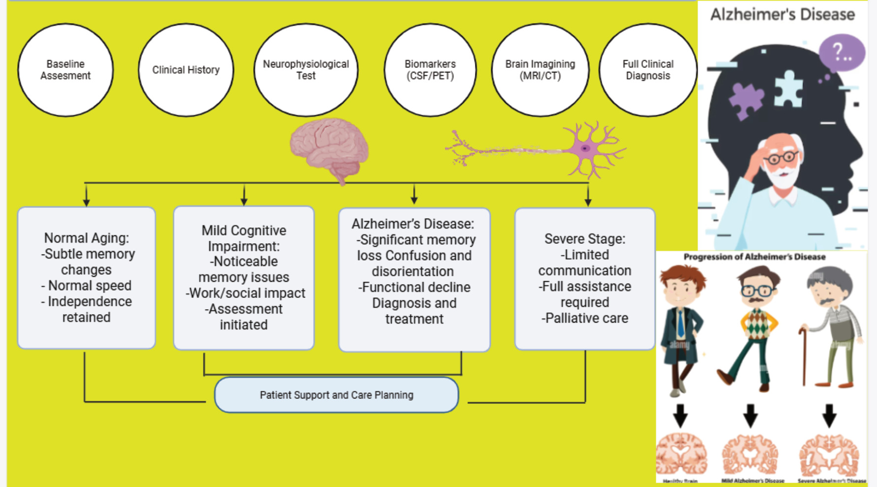
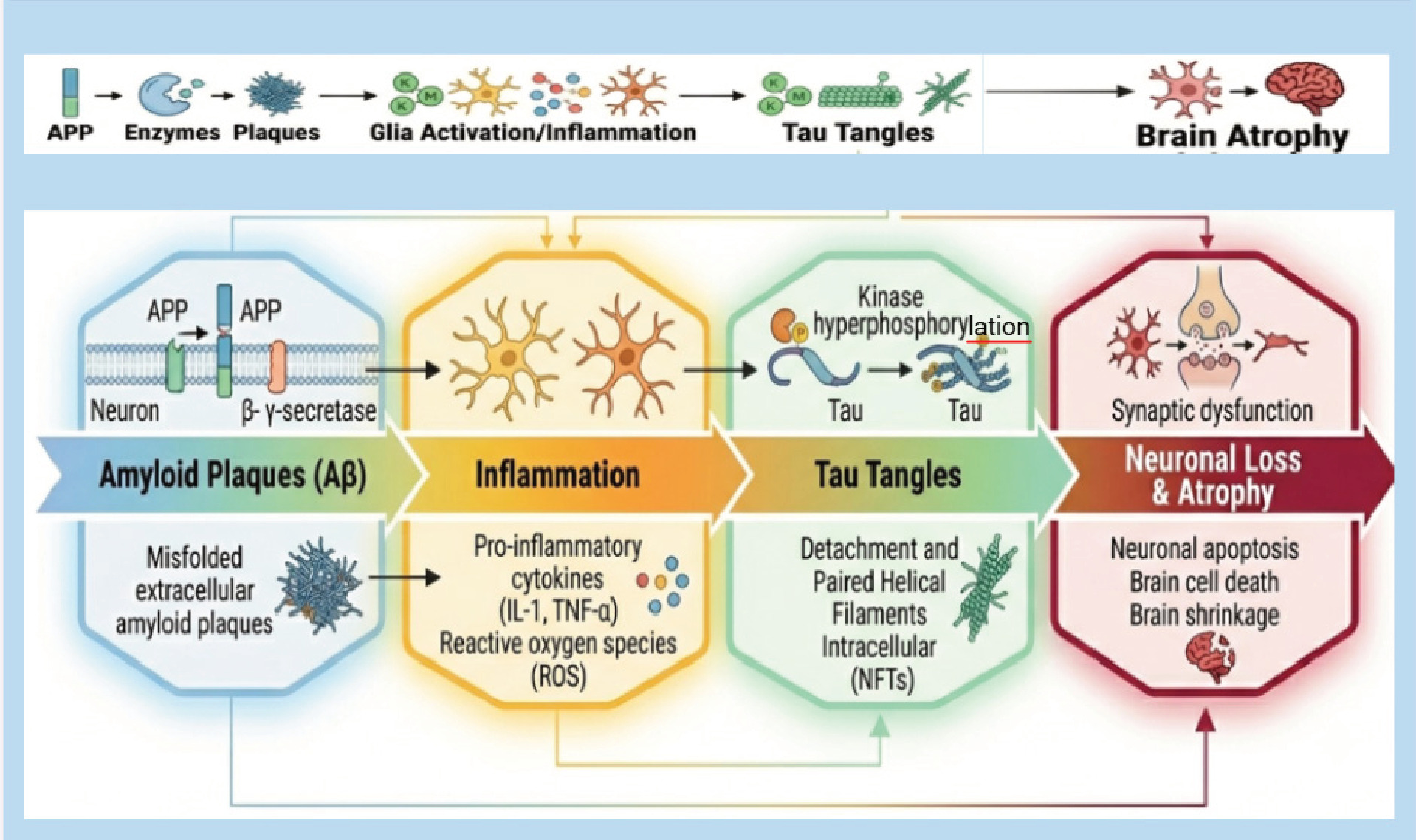
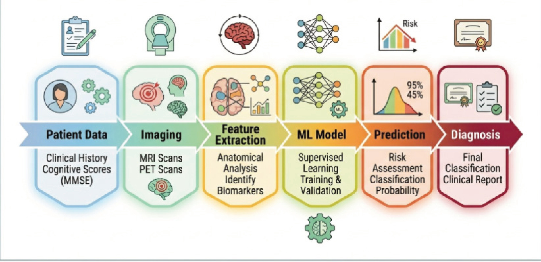
    An example of progressive neurological disease is the so-called Alzheimer's Disease (AD). Alzheimer's disease is characterized by such clinical manifestations as low level of cognitive function, lack of memory, strange behaviour, and inability to perform daily routine on one's own . One can claim that Alzheimer's disease is one of the most common causes of dementia in the world. Furthermore, this disease affects the elderly population only. The main characteristics of Alzheimer's disease are the presence of Beta-Amyloid (Aβ) plaques in extracellular space and neurofibrillary tangles of hyperphosphorylated tau protein inside the neurons . Due to these characteristics, neurons start working improperly, synapses get destroyed, and finally the brain suffer from atrophy. The disease has become a significant public health issue due to the fast-growing number of people who have reached old age. In terms of the recent global statistics, millions of people around the world suffer from dementia, with Alzheimer's disease comprising about 60-70% of all these cases . The number of people suffering from this disease will grow drastically over the coming years due to the increased life expectancy rates. The growing prevalence of the disease not only affects its patients but also creates a great pressure on the healthcare system, families of these people, and economies. The growing prevalence rate of this disease leads to an increased level of expenditure on health care related to treatment, care, and hospitalization. In spite of the remarkable progress made in medical science and the research being done on neuroscience, the diagnosis of Alzheimer's disease still poses a big problem . The conventional methods of diagnosing the illness usually employ clinical assessment, neuropsychological tests, cognitive testing, and analysis of the patient's history. But most of the time, such techniques detect the presence of Alzheimer's only when there are already some signs of the disease, which is usually at an advanced stage of the pathology. Neurodegeneration can start long before any clinical signs are observed . Also, there is the possibility that traditional diagnostic procedures may suffer from subjectivity as well as variation in different medical practitioners. This is because cognitive testing can lead to inaccurate diagnosis in some instances due to the influence of certain conditions like education level, age, emotional state, and neurologic conditions. In addition to this, neuroimaging techniques and laboratory tests, although helpful, may involve specialized skills as well as costs. Differentiating between Alzheimer's disease and other types of dementia or age-related cognitive impairment can also be hard in certain cases. These challenges bring out the need for effective diagnostic procedures. The early diagnosis of Alzheimer's disease has received much attention due to various benefits that arise from the condition being identified early. The early identification of the changes that happen within the body before neurons suffer any damage may help in making important decisions clinically, and as a result, delay the disease progression, thereby maintaining brain functioning for a prolonged period of time. Early diagnosis will also be useful for counselling the patients and their caregivers, changing lifestyles, and other things. Additionally, early identification of individuals at high risk of the disease will help in enrolling such people in clinical trials that seek to develop disease-modifying drugs . There has been increasing importance of using biomarkers in the field of diagnosing Alzheimer's disease during the last few years. These are biological markers that indicate the presence of any biological activity going on inside the body. In case of Alzheimer's disease, these biomarkers may include CSF biomarkers, blood biomarkers, genetic biomarkers, and neuroimaging biomarkers concerning Amyloid beta deposition, tau protein pathology, neuronal injury, and brain changes. The development of neuroimaging techniques including MRI and PET scans has made it possible to identify brain changes because of this disease easily (Miesr et al., 2015). On the other hand, the fast progress in the field of Artificial Intelligence (AI) has brought significant changes in many aspects of health care such as medical imaging and disease forecasting. Various AI techniques, especially Machine Learning (ML) and Deep Learning (DL), have demonstrated a great potential in analysis of big and complex data sets with extremely high accuracy. In combination with biomarkers and neuroimages, AI methods can help find some hidden patterns and anomalies that cannot be discovered by means of traditional data processing methods. It becomes possible to use AI to conduct automated data feature extraction, classification, forecasting, and risk evaluation, thus facilitating quicker and more accurate diagnosis for clinicians . Considering the rising significance of Artificial Intelligence technology and biomarker studies, it is essential to have a comprehensive knowledge base about the latest advances in this highly dynamic area. It is the main goal of the present review to provide a thorough analysis of Artificial Intelligence-based biomarkers and neuroimaging techniques for the early detection and diagnosis of Alzheimer's disease. The present review is aimed at discussing various types of biomarkers, neuroimaging techniques, Artificial Intelligence methodologies used in Alzheimer's disease studies, and their clinical importance. Moreover, the challenges and perspectives related to the application of Artificial Intelligence techniques in clinical practice in Alzheimer's disease studies are emphasized as shown in (Figure 1) . Pathophysiology of Alzheimer's Disease Alzheimer's Disease (AD) is a multifactorial progressive neurodegenerative disease caused by some pathologic factors that are capable of causing injury to neurons and their cognitive functioning. Even though the etiologic cause of the disease has not been identified yet, there are certain physiological factors which have been found to be associated with AD pathology. The pathophysiology of Alzheimer's disease involves accumulation of amyloid beta peptide, dysfunctional activity of tau protein, neuroinflammation, vulnerability to AD genetically and cellular damage due to oxidative stress. These factors interact with each other and eventually lead to synaptic dysfunction, neuronal death, and changes in brain structure . Amyloid-Beta Hypothesis For a long period, amyloid-beta theory has been viewed as one of the most credible theories concerning the origin of Alzheimer's disease. As per the theory, the emergence of Alzheimer's disease is caused by the incorrect APP processing. The normal physiological function of the protein includes processes of neuron regeneration and growth. In normal conditions, the protein is processed through non-amyloidogenic mechanism. In contrast, the APP processing by β-and γ-secretase under the conditions of Alzheimer's disease results in the formation of Aβ peptides, namely Aβ40 and Aβ42 . All the peptides are dangerous, but the most dangerous is Aβ42 because it tends to aggregate in insoluble extracellular plaques. The presence of amyloid-beta plaques affects the work of neurons and initiates several negative physiological mechanisms. Namely, the formation of amyloid-beta interrupts the synaptic processes, disrupts the balance of calcium and provokes inflammation. Moreover, amyloid-beta plaques cause the oxidation and death of neurons. While the amyloid-beta hypothesis has helped us gain more insight into the pathophysiology of Alzheimer's disease, several findings have shown that the presence of amyloid alone cannot explain the progression of Alzheimer's disease. Studies have suggested that the amyloid pathology might work along with other factors like tau pathology and inflammation in Alzheimer's disease . Tau Protein Abnormalities Abnormally modified tau protein constitutes another distinguishing characteristic of Alzheimer's disease. Tau protein refers to microtubule protein, whose major function is to provide stability to the structure of nerve cells and help in intracellular transport of these nerve cells. Normally, the tau protein associates with microtubules to ensure proper functioning of neurons (Jhonson et al., 2004). However, in the case of patients with Alzheimer's disease, there develops an abnormal situation in terms of hyperphosphorylation of tau proteins. As a result, such proteins become dysfunctional and get dissociated from the microtubules to form intracellular aggregates, known as Neurofibrillary Tangles (NFTs). Neurofibrillary tangle formation is another major factor in neuronal degeneration and brain shrinkage. Research has shown that the occurrence and evolution of the pathological presence of tau are related to a significant extent with cognitive decline and the degree of the disease itself. In contrast to the occurrence of amyloid deposits that can develop many years before the onset of symptoms, the pathology related to the presence of tau is often linked to memory loss and functional decline . Neuroinflammation Another crucial factor involved in the development of Alzheimer's disease is neuroinflammation. Neuroinflammation is associated with the functioning of microglia and astrocyte cells. They have a protective role in the central nervous system when functioning normally and maintain the balance in the brain since they eliminate the dead cells and toxins. Nevertheless, prolonged activation of the immune cells leads to chronic inflammation. The formation of amyloid-beta plaques and neurons' damage stimulates microglia activation and secretion of pro-inflammatory agents, for example, cytokines, chemokines, and reactive oxygen species. While an acute inflammatory process gives protection, chronic inflammation is dangerous and causes damage to neurons. Chronic neuroinflammation is accompanied by an increase in the amount of amyloid, hyperphosphorylation of tau proteins, and neurons' death. Also, signaling in the presence of chronic inflammation negatively influences the synapses and promotes cognitive impairment. Scientific research shows that neuroinflammation is not only the effect but also the component of Alzheimer's disease . Genetic Factors Moreover, it is obvious that genes contribute to an increased susceptibility towards Alzheimer's. This either is in cases of familial (early onset) form, of which few, which involves a mutation in a specific gene, namely the Amyloid Precursor Protein (APP) gene, as well as in Presenilin-1 (PSEN1) or Presenilin-2 (PSEN2) genes which may be involved in alteration of the processing of amyloid peptides and can lead to higher formation of amyloid beta species which in the pathogenic one's cause diseases. Most patients, nevertheless, suffer from AD with a late onset and sporadically influenced by many factors (genes, environmental conditions and life style) and risk factors . Oxidative Stress There is another key player in Alzheimer's: oxidative stress. In a nutshell, Oxidative Stress happens if the cellular antioxidants balance their defensive abilities by generating over-the-top free radicals . The brain's metabolic rate is very high; this is one thing the oxidative protection cannot match. That means Alzheimer-suffering patients generally face a bigger danger of suffering from it. The free radicals have a way to damage the lipids, protein structure, even DNA let alone membranes then you just know nothing can work right, and the Neurons have been injured or killed. Scientists think that one cause of Alzheimer's is that amyloid plaques build up and mitochondria malfunction; this leads to the production of so much free radicals that more Amyloid can stick and tau-protein build up and eventually, the Neurons are injured. These processes together promote inflammation of brain and impairment of neurons' connections that leads to cell death. It makes sense why the scientific community has to focus so much attention on decreasing of oxidative damage. To put it in a nutshell, Alzheimer's develops due to several co-dependent biological process that make Neurons die, and result in a loss of cognitive function. This knowledge can bring about new biomarkers and have been extensively researched to bring about innovative AI solutions for a more accurate diagnostic tool as well as for individual therapeutic management strategies as shown in (Figure 2) . Biomarkers in Alzheimer's Disease In the diagnosis, monitoring, and characterization of Alzheimer's Disease (AD), the application of biomarkers has increasingly become indispensable. A biomarker is defined as an objective measure used to study and indicate normal biologic processes, the action of drugs, or responses to therapeutic treatment of disease . Because the process that leads to Alzheimer's disease begins a decade or more before it is clinically evident, researchers are exploring biomarkers that can detect AD earlier and support a clinician's efforts to discriminate it from other neurodegenerative conditions. Discoveries over the last few years in molecular biology, genetics, and neuroimaging have characterized multiple biomarkers of amyloid accumulation, tau pathology, neuron degeneration, and architectural brain abnormalities that may enable a new generation of biomarkers that target a range of AD-associated phenomena in CSF, blood, gene expression, and brain imaging . Combined approaches to biomarkers have shown tremendous promise for more precise and earlier detection. Cerebrospinal Fluid Biomarkers Alzheimer disease CSF biomarkers: One of the most studied and clinically tested CSF biomarkers used for the diagnosis of Alzheimer's Disease. Cerebrospinal Fluid (CSF) surrounds your brain and spinal cord which means abnormalities occurring in the central nervous system can manifest within the fluid. By measuring particular proteins in CSF samples, it is possible to get an idea of the specific underlying pathologies of the brain occurring throughout Alzheimer's . Amyloid-beta 42 (Aβ42) Amyloid-beta 42 (A42), a marker of disease which may predict the future risk of dementia Amyloid beta 42 is a substance in Cerebrospinal Fluid (CSF), which is strongly suspected to contribute to the disease processes occurring in the brain (Hannson et al., 2019). A42 is created by the enzymatic cutting of amyloid precursor protein, a large protein precursor, and is a strongly aggregated and more readily deposited protein product compared to the less soluble variantA40. In individuals with Alzheimer's disease CSF concentration of A42 levels tend to be lowered, as the more amyloid beta peptides are deposited in brain tissues in the form of amyloid plaques the levels in CSF decline. Lower levels may even be observed for some years before the clinical manifestation of the disease and can be a useful tool for prediction. The reduced levels of CSF A42 have shown promising results in numerous studies when distinguishing between Alzheimer's disease patients and cognitively healthy controls or patients with other types of dementia. Because A42 alone, however, has its limitation, A42 is generally measured in combination with some markers based on tau . Total Tau (t-Tau) Tau proteins are essential structural and transport molecules of the microtubules in neurons, responsible for maintaining cell integrity. Tau proteins are released into the CSF as a consequence of neuronal damage . Total Tau (t-Tau) Total tau represents the sum concentration of all tau protein in the CSF and can be an indication of neurodegeneration and neuronal damage. Increased total tau levels has frequently been described in Alzheimer's Disease patients and are generally regarded as proportional to neuronal damage in this population . It does not distinguish between Alzheimer's pathology from other sources of neuron damage, unlike the amyloid beta tests, and therefore, while it provides valuable data as part of a biomarker combination, it would not by itself diagnosis the condition, although it can offer a good indicator of disease progression and severity. Phosphorylated Tau (p-Tau) This indicates that p-Tau represents a more specific marker of Alzheimer's disease pathology. In AD, tau proteins are abnormal hyperphosphorylated, forming neurofibrillary tangles in neurons. The level of phosphorylated tau in CSF is well correlated with the underlying tau pathology and formation of neurofibrillary tangles. It also appears more specific to AD than does total tau, with promising evidence in differentiating Alzheimer's from other forms of neurodegenerative diseases. A panel composed of lower A42 and higher t-Tau/p-Tau has already been one of the most utilized profiles of AD biomarkers . Blood Biomarkers However, while the biomarkers found in CSF can offer much-needed insights, CSF is accessed via lumbar puncture which can be inconvenient and might not be suitable for large-scale screening. Hence, much effort has been made into blood biomarkers that are easy and cost-effective to obtain by simply using simple blood test methods. Plasma Tau proteins, a new class of biomarkers, hold promise for Alzheimer's diagnosis and follow-up. Development of ultrasensitive tau detection methods allows for the quantification of low plasma concentrations. Several research findings indicate that increased plasma tau is related to clinical symptoms and disease severity, with specific phospho-forms such as p-tau181 and p-tau217 showing significant diagnostic ability in a blood test for Alzheimer's . This has implications for population-based screening programs to facilitate early detection of at-risk individuals. Additional validation studies, however, need to be performed before it can be implemented in routine practice. Neurofilament Light Chain (NfL) NfL a biomarker of neurodegeneration The NfL, one component of the structure of the axone, can pass into our body fluid when neuronal lesions or neuron degeneration occurs . High blood NfL concentrations in patients diagnosed of many types of neurodegenerative disease, such as the Alzheimer's disease, have been reported. Higher plasma concentrations are markers of an active neuronal loss and may correlate with clinical decline and progressiveness of the disease. While NfL is not a disease specific biomarker, it is a good general marker for neurodegeneration which may be useful for tracking patients. This marker together with others may have a higher value for diagnosis and prediction. Genetic Biomarkers The understanding of diseases risk including the predisposition to the AD can help find affected individual with a greater tendency. Genetic predisposition is an essential determinant of Alzheimer's Disease, both the familial and the sporadic forms . APOE ε4 In relation to the development of late-onset Alzheimer's disease, APOE is a genetically significant contributing factor, having the strongest established linkage. In humans, three major variations (APOE 2, 3 and 4 allele) of the APOE gene occur, among which is the APOE4 allele, which is highly correlated with disease likelihood . In relation to development of Alzheimer's, one copy of APOE 4 appears to confer risk, whilst a second copy has a high probability of presenting an even greater risk of development, and potential earlier age of onset. As one APOE 4 copy does not directly lead to development, but contributes by acting on various mechanisms contributing to the pathogenesis of Alzheimer's namely amyloid metabolism, deposition and repair, APOE genetic profiling is routinely considered for in Alzheimer's research . Gene Mutations Mutations in specific genes have also been identified with the cause of Alzheimer's disease. APP gene mutation, PSEN1 gene mutation, and PSEN2 gene mutation are usually linked with the earliest occurring forms of Alzheimer's disease . These gene mutations influence amyloid metabolism and thus increase the number of damaging amyloid-beta peptide. However, the number of people suffering from familial Alzheimer's disease are smaller compared with people from the non-familial Alzheimer's cases. Imaging Biomarkers An introduction to brain biomarkers used to measure structural changes during the progression of Alzheimer's disease. These imaging techniques are non-invasive tools that allow physicians to capture the structural, functional, and molecular changes in the brain that occur with this form of neurodegenerative dementia . The different types of brain biomarkers for the study and analysis of Alzheimer's' are discussed below. Magnetic Resonance Imaging (MRI) is widely used for evaluating structural changes associated with Alzheimer's disease. MRI provides high-resolution images of brain tissues and allows the assessment of regional brain atrophy. One of the most important findings observed in Alzheimer's disease is hippocampal atrophy, which is strongly associated with memory impairment . MRI can also identify cortical thinning, ventricular enlargement, and reductions in gray matter volume. Additionally, advanced MRI techniques such as functional MRI and diffusion tensor imaging can provide information regarding neuronal connectivity and functional alterations . Positron Emission Tomography (PET) With the use of advanced tracer technology, Positron Emission Tomography (PET) may assess not only anatomic information but also some changes of molecular or biologic nature in the brain such as detection of both tau pathology and/or Amyloid-Beta (Abeta) plaque deposition . Amyloid PET could be able to demonstrate accumulation of Abeta in a relatively early stage, whereas FDG-PET may asses regional metabolism which is depressed in the regions afflicted with Alzheimer Disease (AD) . Although its utility is evident, PET is relatively costly, and not readily available. Computed Tomography (CT) imaging provides cross-sectional views of the brain anatomy, and may also be utilized for assessing brain atrophy, as well as to rule out other potential neurological diseases . The CT imaging has been shown to be less sensitive than MR imaging for the subtlest abnormalities encountered in the very earliest stages of AD. CT imaging may be more readily available, inexpensive, and acquisition of images can be performed quickly in many centers, supporting an important role of CT imaging as a tool that helps a physician identifying structural abnormalities and supporting differential diagnosis in clinical practice. In summary, biomarker techniques have revolutionized the diagnosis and characterization of AD through improved early and accurate detection of pathological features of the disease, whereas combination of several biomarker modalities with Artificial Intelligence based approaches will further boost diagnosis specificity and will support tailored therapies for the AD as shown in (Table 1), . Artificial Intelligence in Alzheimer's Diagnosis With the continuous development of biomedical data resources, computing technologies, and machine learning algorithms, Artificial Intelligence (AI) has become a prominent technology, which has made a significant contribution to the diagnosis and management of neurological diseases such as Alzheimer's Disease (AD) . The application of AI technologies in Alzheimer's disease has shown its capacity to enhance diagnostic efficiency, monitor the progression of underlying pathology in early stages, and implement personalized treatment approaches. Alzheimer's disease diagnosis usually involves the application of imaging examination, cognitive tests, biochemical and immunological indicators and genetic tests in clinic examination. However, traditional diagnosis method cannot detect pathological change in early stage well. By learning from a lot of different kinds of biomedical data including scans, biomarkers, blood markers, genes and patients' status, AI techniques have the capacity to mine concealed regularities hidden in those high-dimension data and help to make an early prediction. Several AI methodologies can be adopted to apply to the AD diagnosis including machine learning, deep learning, predictive analytics and clinical decision-support systems . These are becoming a central part of the precision medicine applications. Machine Learning is a subfield of artificial intelligence which permits the systems to achieve knowledge of data with none explicitly programming, to form predictions and identify knowledge patterns from knowledge. The machine learning algorithms area unit significantly used in Alzheimer's disease analysis to classify patient into specific classes, discover the new Alzheimer's disease biomarkers, and predict progression of disease . The structure of the Machine Learning models comprises of data preprocessing, feature extraction, training and classification, respectively. The clinical information, neuroimage feature, genetic data and biomarker levels are typically used as input. Machine learning models then capture the relationship in the dataset and then apply these trends to classifying an individual as normal, MCI or Alzheimer's disease. Advantages are: Accuracy, less human bias, Handling huge dimensionality datasets. Several ML techniques are successful for the diagnosis of Alzheimer disease . Random Forest is an ensemble machine learning that uses a set of decision trees to improve prediction accuracy, because not using one but multiple trees, but several individual ones and to combine results so to perform final prediction. Random Forest has been applied in study on AD because they work fine for the high number and the complex interactions of features available for AD-related datasets like the ones in neuroimaging, for biomarkers, in demographic information and for genetic data (Lia et al., 2022). These models offer information about feature importance which make researchers find the most critical biomarkers for disease development. The fact that it has built-in methods for missing values and a way to reduce the possibility of over-fitting contribute to make Random Forest highly attractive in clinical context. It has already been shown that these models have satisfactory accuracy on distinguishing AD from healthy participants as well as from those with mild cognitive impairment in AD classification. Support Vector Machine (SVM) In the field of Alzheimer's research, SVM is one of the frequently used supervised ML techniques . It performs classification of various groups by identifying the boundary line or the hyper-plane which differentiates the various groups and tries to increase the difference between the various groups to improve the classification accuracy. As SVM are particularly successful for high-dimensional biological information from MR scans and biomarker-based data set. Hence, a lot of researches used SVM techniques on classification of healthy subject from patient with mild Alzheimer's to severe disease and to estimate progression of disease. Research shows the efficiency in distinguishing between the different types of AD. Another main strength of SVM is that it is one of the few techniques used even with smaller samples of patients with promising efficiency, however the parameter, and the kernel selection for better results need optimization of the algorithm for classification. Decision Trees Another popular algorithm that falls under this category is the usage of decision trees in disease classification. Decision trees, as the name itself suggests, use tree-like models to arrange data that leads to final classification outcomes, with a split of datasets based on selected characteristics of data, repeating recursively, until finally a disease is diagnosed. The data sets can vary widely, in the Alzheimer's disease context it can consist of biomarker results, images, etc. This type of classification algorithm has certain advantages due to the clarity and easy to follow nature of visual representation. Decision trees also present the advantage to interpret, easily, the results leading to a specific classification thus enabling clinical decision . However, single decision trees are often observed to be unstable and perform poor by causing over fitting, hence many ensemble techniques including random forests incorporate them. Deep Learning Machine learning and artificial intelligence are both fields that contain machine learning technologies and artificial intelligence techniques. Machine learning has, more recently developed in more details into a concept, called deep learning. The deep learning is an artificial algorithm by using information to perform different patterns which contains machine learning, etc... It represents itself with deep stacked layers of artificial neural network for pattern recognize, in order to learn representation directly from data without the previous feature extracted . This method can find some pattern through huge amount data and help a doctor when dealing Alzheimer disease. Convolutional Neural Networks (CNN) Convolutional Neural Networks (CNNs), a popular category of deep neural architectures, are frequently applied to the area of medical image analysis. CNN-based techniques are well-suited for the analysis of pictorial data that occurs frequently with MRI, PET, and CT imagery . Image data in medical applications can undergo pattern detection via CNN's, which will utilize these "filters to extract feature representations automatically". In instances such as Alzheimer's detection, there are various features that the CNN can recognize: "hippocampal volume, cortical thickness, and pattern of organization among brain tissues". Researchers are successfully designing CNN algorithms for medical data as studies have been published showing high diagnostic precision in distinguishing Alzheimer's from normal subjects, and some that identify individuals who have Mild Cognitive Impairment (MCI) and may likely decline . Such methods will further benefit from fewer labour-intensive feature engineering processes. Recurrent Neural Networks (RNN) A deep learning approach to longitudinal analysis of Alzheimer's Disease (AD) is progressive. Patients often have medical records and clinical assessments taken on several occasions during their life span that accumulate information about progression of disease. Recurrent Neural Network (RNN) is a category of deep learning methods used to predict output values using previously predicted or previously calculated outcomes. RNN model can be trained and is able to capture the relationship of input output variables and can utilize that information to analyze temporal progression or future events . In case of studying medical history, it may be important to include time-series data of some variables such as multiple evaluations of cognitive score over time, levels of AD biological markers, patient's status throughout life-time and clinical records. RNN can utilize that information by including memory cells in the deep learning structure, analyze trends in disease over time and forecast clinical consequences or predicts future health states (Patharker et al., 2024). RNN was successfully utilized to predict future decline of AD on the base of clinical, biomedical data. Long Short-Term Memory (LSTM) network is a type of RNN that can effectively capture long-range interactions in sequences. LSTM was employed by several recent study of Alzheimer to predict disease severity or stage . Predictive Analytics involve using machine learning algorithms, historical data, and statistical methods to predict the likelihood of future events. Scientists have developed algorithms for predicting which individuals are most likely to develop Alzheimer's disease before they develop more obvious cognitive problems . Alzheimer's research centers predict risk by combining patient data sources including health history and biomarkers from clinical data, genetics, and medical images of brains to assess disease risk and rate of progression. Models may be used to predict who among those with MCI (Mild Cognitive Impairment) may develop Alzheimer's. Having risk scores means patients can participate in preventive therapies and take Lifestyle measures to try to keep brains healthier, which would mean having more healthy neurons available when treatments are prescribed. Predictive models may also help trial planners choose suitable participants to receive innovative therapies . The systems that employ Artificial Intelligence can also identify populations needing extra observation and early healthcare services. Clinical Applications With the introduction of AI into clinical care, exciting opportunities exist for enhancing the accuracy of the diagnosis of Alzheimer's disease, and its management. As a clinical decision support tool, an AI may help clinicians make better decisions using various pieces of patient data. For instance, neuroimaging techniques, such as MR and PET imaging, have traditionally been vital for diagnosing neurological diseases, and AI analysis may accelerate this diagnostic process through the automated detection of abnormalities . Moreover, AI offers further possibilities, such as processing biomarker data. If multiple biomarkers are coupled with imaging data and genetic information, an AI model can provide a tailored approach to disease diagnosis and prediction. The future of AI also offers potential applications in remote patient monitoring and digital health systems, utilizing technology such as wearables to perform constant real-time cognitive and behavioural assessments. However, a number of obstacles may hinder clinical adoption. Future clinical adoption of an AI decision support will be dependent upon overcoming obstacles concerning data quality, model interpretability, privacy, and the ethical implications. Big and unbiased data needs to be made available to facilitate generalisation of such AI models. Transparency and the explanation of how the algorithm comes to conclusion should also be in place so that clinicians will trusts such AI model in decision making and utilize AI in assisting clinical processes. AI is currently at a crucial juncture in Alzheimer's diagnosis and with the combination of techniques such as machine learning, deep learning, predictive modeling, AI has huge potential in early detection of diseases and enabling highly individualized Alzheimer's patient care . Future developments will allow AI to play an even greater role in Alzheimer's disease, as shown in (Figure 3). Neuroimaging Approaches Alzheimer disease diagnosis and understanding with neuroimaging Alzheimer Disease (AD) diagnosis, and pathogenesis evaluation and understanding has substantially relied on different neuroimaging approaches . These methods allow us to probe into alterations of the structure, function, and molecules within the AD affected brain. Alzheimer pathology can begin in the brain many years prior to clinical symptom onset, and the imaging studies become more useful in an early detection of the disease and to follow disease progression with time. The improvements of imaging technique render us able to visualize the abnormalities that occur during AD such as the deposition of amyloid plaque, degeneration of neuronal cell, alternation in neuronal connection, and change in the brain metabolism and functionality. Different kinds of cognitive tests and clinical observation might be unable to perceive minimal alterations in the disease in the early phase of Alzheimer's disease while neuroimaging examination provides a quantitative measure to confirm the diagnosis, to characterize the pathogenesis and importantly to develop AI-driven disease risk prediction and identification of biomarkers of AD. The main types of neuroimaging techniques used to study AD are: Magnetic Resonance Imaging (MRI) MRI is one of the most common neuroimaging techniques used to examine structural brain changes found in Alzheimer's disease. It relies on strong magnetic fields and radiofrequency pulses to produce detailed image scans of the brain's structures without the use of ionizing radiation. Due to its good spatial resolution and soft tissue contrast, MRI has become a valuable tool in the assessment of neurodegenerative conditions. Changes found in MRI of the Alzheimer's brain include: Atrophy of the hippocampus (a structure critical for learning and memory that is extremely vulnerable to Alzheimer's-related cell loss). The decrease in size of the hippocampus is one of the first signs of the condition on brain scans. The progressive reduction of the thickness of the outer part of the cortex (Gray matter). Progressive dilatation of brain ventricles. Reduction of overall brain volume. It's not limited to changes in the hippocampus; Alzheimer's is also typically associated with shrinkage in regions such as the frontal, temporal, and parietal lobes. Changes in structure can correspond with the worsening of Alzheimer's as cognitive symptoms progress. Other methods MRI is being explored more for its use as an imaging technique for identifying and characterizing other changes occurring in Alzheimer's, such as: Using Diffusion Tensor Imaging (DTI), a special kind of MRI technique used for examining water diffusion in the body that may reveal white matter abnormalities in Alzheimer's . Functional MRIs are also being studied in an attempt to identify Alzheimer-related differences in brain activity patterns. MRI has advantages that include being non-invasive, using no radiation, providing good anatomical images and having the capacity to distinguish between various tissues in the body. Its limitations include its cost, time-consuming procedures, and its failure to adequately show evidence of early disease onset in Alzheimer's. Positron Emission Tomography (PET) The Positron Emission Tomography (PET) scan is a useful tool in assessing metabolic and cellular functions in the body's organs such as the brain . For AD the test takes radioactive tracer substances which in turn build up into regions with the particular disease being visualized to determine abnormalities with regard to Alzheimer's' . 1) Assessment of the amount of beta-amyloid being deposited is being done with special PET scan imaging. Amyloid in brain tissues deposits and builds up even a number of years before there are any symptoms related to Alzheimer's. With special tracer for amyloid PET imaging will show the area of plaque build-up within brain cells , which allows one to observe at an early time that one has increased risk factors or if they already have AD without outward signs yet. 2) FDG-PET scanning. This is another used PET scan for AD. PET can tell the activity level of glucose metabolism in the brain's cells. Glucose in brain's neurons fuels with high energy level to provide brain cells with its need of glucose to be carried out properly. A decrease of metabolic activities and consumption rate of glucose may be an indication of loss of brain functioning and or disease progression. It has been shown that patients that has suffered Alzheimer's, there is often a decline in metabolism in either areas of their parietal or temporal cortex of their brains. This decrease can often be detected during the progression stage of Alzheimer . PET has now been used to examine tau pathology through imaging of the abnormal tangle formed from tau in Alzheimer's disease, which will help for better understanding from both the assessment of both beta-amyloid and tau pathologies simultaneously in the disease process . However, due to expense, accessibility to special facility and radioactive exposure PET cannot be widely available. Functional Magnetic Resonance Imaging (fMRI) is a high-tech method of imaging brain activity by observing changes in oxygen levels in the blood. Functional MRI differs from other methods of imaging the brain that the other images mainly observe for anatomical alterations like MRI and not changes of activity. Since the activity of neurons within the brain demands a supply of blood, when active blood flows throughout, supplying the necessary oxygen and nutrients and increasing the oxygen level in the surrounding tissues of brain. The Blood Oxygen Level-Dependent (BOLD) signal generated by the brain from fMRI is observed and presented on images showing brain activity. The studies in Alzheimer disease research have utilized fMRI widely . These studies were aimed at examining the change in functionality and network connections of the brains of people. It has been proved through the fMRI study that the communication of signals in between different parts of human brain is not that consistent in people suffering with Alzheimer disease, especially the sections of brain that are involved in different cognitive tasks. The most talked about brain network DMN, which consists of the brain sections responsible for cognitive functions as well as memory, shows abnormal brain activity in Alzheimer patients, even at an earlier stage. Additionally, functional MRI images provide us an overview of compensatory responses, in the event that one brain area enhances functioning to compensate the loss of functionality. The information provided by the technique has very importance in study and understanding about brain diseases. Functionality, is what that concerns a number of scientists studying such diseases, so non-invasive, as this technique is, is it worth applying. Unfortunately, it may get hard sometimes to interpret the signals that generate out of this process of brain examination because of complexity. Electroencephalography (EEG) is a non-invasive neurophysiological procedure performed for the recordation of electrical activity generated by neurons (or brain cells) . EEG allows us to assess the activity of the brain over time with use of electrodes that have been placed on the outer skull region. Different than MRI, and PET imaging techniques EEG has excellent temporal resolution, allowing for rapid assessment of neural dysfunction in real-time. In recent years, the interest has grown about usage of EEG, since it is cheap, mobile, allows examination of the neural abnormalities associated with Alzheimer's Disease, and has the ability to define alterations in EEG which can be unique to AD. In the context of AD, there have been evidences showing characteristic patterns of alteration in EEG including slowing of the brain wave activity, diminution of higher frequency waves, and increased low frequency activity. Such patterns indicate defective neuronal interaction and impaired cognition. EEG is also used to examine the functional interconnectivity of brain regions as well as changes in neural networks related to disease progression . A variety of EEG-derived markers have shown evidence as useful indicators for early diagnosis and forecast of cognitive deterioration. Moreover, the usage of EEG for Alzheimer's' classification by applying Artificial Intelligence is now a subject of interest and several studies demonstrated good performance achieved by applying various artificial intelligent algorithms like machine learning and deep learning for that task. The main drawbacks of EEG include relatively low special resolution compared with PET and MRI imaging and susceptibility of EEG signals to artifact and external noise. In conclusion, neuroimaging is providing new avenue in diagnostics of AD with visualization of various pathological changes (molecular, structural and functional) in the brain in addition to various forms of AI integrated analysis and biomarker-based techniques, as shown in (Table 2).

    CURRENT CHALLENGES AND LIMITATIONS

    While Artificial Intelligence (AI), biomarker technologies, and neuroimaging have shown tremendous promise in enabling early and accurate diagnosis of Alzheimer's Disease (AD) and improved management outcomes for patients, there are several challenges and obstacles to its broad adoption and application in clinical settings. Efforts toward developing sophisticated machine learning algorithms, deep learning systems, and biomarker-integrated diagnostic tools have been quite successful, but the integration of these technologies from the research laboratory into standard clinical use has proved to be arduous. Many aspects-data utilization and acquisition, model reliability and predictive validity, ethical issues, costs and reimbursement, and the need for validation in clinical practice-are persistent issues in the development of effective and widely deployable AI tools for AD diagnostics . Successfully addressing these problems will provide the necessary technology framework that allows health care professionals to accurately and efficiently make important decisions and provide timely interventions for the benefit of patients. Small Datasets A Significant Challenge for AD diagnosis models -The paucity of sufficient training datasets for developing Artificial Intelligence models for diagnosing Alzheimer's Disease (AD), an enormous and diverse data is an unavoidable requisite. In general, large amounts of high-quality training data are needed to build robust deep learning AI models and optimize their performance. Nevertheless, collecting large training data in Alzheimer's research could be rather difficult to comply, due to a host of clinical and/or experimental restrictions. The size of Neuroimaging based patient studies usually limited by cost, scan duration (MRI and PET scans can be costly) and the procedures to obtain CSF and longitudinal data can be considered to be invasive, this is one reason why patients were less readily enrolled in AD studies . Datasets used, may vary due to different participating hospitals or sites; patients may show variation in demographic features, imaging methods, and clinical assessments across differing clinical practices or sites and thus this would potentially affect generalization capabilities. Limited sized data increases the probability of generating sampling bias so, AI model built from small sized sample is capable to perform very well at experiments, but the model may not perform well at clinical practices. Overfitting is another crucial challenge. In machine and deep learning for Alzheimer's disease, overfitting happens when an AI model becomes so highly tuned to the specific training set and learn to capture irrelevant patterns and noise instead of generalizable characteristics of the disease pathology. The model is good at training, but when it is used on unseen and new data. This type of issue is really serious in medicine since usually training datasets tend to be relatively small and of high dimensions. For instance, many neuroimaging datasets can contain thousands of Features from MRI or PET data. Whereas available patient number are still smaller than feature number. There were some methods developed to deal with over-fitting including data augmentation, cross-validation approaches, regularization, ensemble learning. And transfer learning where existing trained model were utilized. Nevertheless, the optimal compromise between complexity of the model and generalization remains to be found. New datasets will give better results . Ethical Issues Ethical Concerns Artificial Intelligence has continued to become more popular across healthcare system use; it has raised ethical considerations about the use of diagnostic AI technologies that collect, use, and store sensitive personal patient information that includes personal medical records, genomic data, biomarker profiling, and neurological imaging results. The protection of patient's confidential information, privacy, and data can compromise patients with any information theft and unauthorized usage that can harm patients and organizations alike. Data governance processes, as well as specific laws to protect the data, are needed. Algorithmic bias is one of the most critical issues faced in using the AI for health-tech development . The AI system learns using training data that represents particular demographic segments if the dataset that AI has been trained upon does not encompass diverse and relevant data-based patterns, there could be a likelihood that AI algorithms develop bias that's expressed in an unequal health-related decision and result to health system disparity. In the case that there is specific racial group training, it might lead to a poorer prediction rate if other ethnicity demographic based models being tested for the same diagnosis. AI's transparency, explainability is another issue. One main area for AI concerns a variety of deep learning systems: that it's usually a 'black box' . Many AI-based deep learning systems for diagnose can be described as 'black boxes' and the underlying decision-making logic might not clear for physicians to understand what is leading their final outcome. Health care providers may be less trust and confidence with the AI diagnoses or predictions if there is any uncertainty with the decision processes, so, the results derived cannot be understood. Lastly, the discussion of informed consent, data ownership and decision liability are other concerns related to AI. Cost Financial factors, too, pose significant obstacles for the adoption of AI-based biomarkers and neuroimaging in AD diagnosis . Training and utilizing an AI system can be extremely costly, demanding massive computing power, specific software and talented individuals to implement and maintain it. In addition, neuroimaging scans such as PET and MRI scans may not be available or affordable in low-resource environments. PET imaging requires specialized equipment as well as radioactive tracers, driving up costs significantly. A variety of biomarker tests, from CSF to genetic tests and novel blood assays, can all contribute to the overall burden of the treatment regimen. Furthermore, many AI-based applications will need continuous upgrades, maintenance and technical support; the financial burden associated with these procedures, and limited resources in many health systems, will most likely result in low-cost diagnostic solutions and tools being the only ones available . Clinical Validation Issues While many of the research studies demonstrate encouraging outcomes, the majority of AI-based diagnostic systems have not yet undergone thorough clinical validation for general use in health care settings. Most research trials for deep learning and machine learning models are conducted in laboratory settings and may not mimic the complexity of an actual clinical setting. Variations between patient cohorts, medical imaging equipment and healthcare delivery may result in varying degrees of model accuracy when the AI systems are implemented into the clinical setting. The accuracy achieved with a training data set often does not transfer to similar accuracies when the algorithms are applied to independent external test data sets. Standardization of the methods of data extraction, data processing, training process, and model evaluation is an additional problem that limits comparison between models. Many of the studies reported thus far use retrospective data, and larger prospective clinical studies conducted on an international multicentre scale and of sufficient follow-up duration is required before the models can be proven reliable and generally applicable. Before deployment into the clinical sphere, many new health care diagnostic technologies also need to satisfy regulatory oversight processes. Agencies need to be convinced about the safety, efficacy, reproducibility, clinical utility and cost-effectiveness of AI based approaches before they can be granted approval for use .

    FUTURE PERSPECTIVES

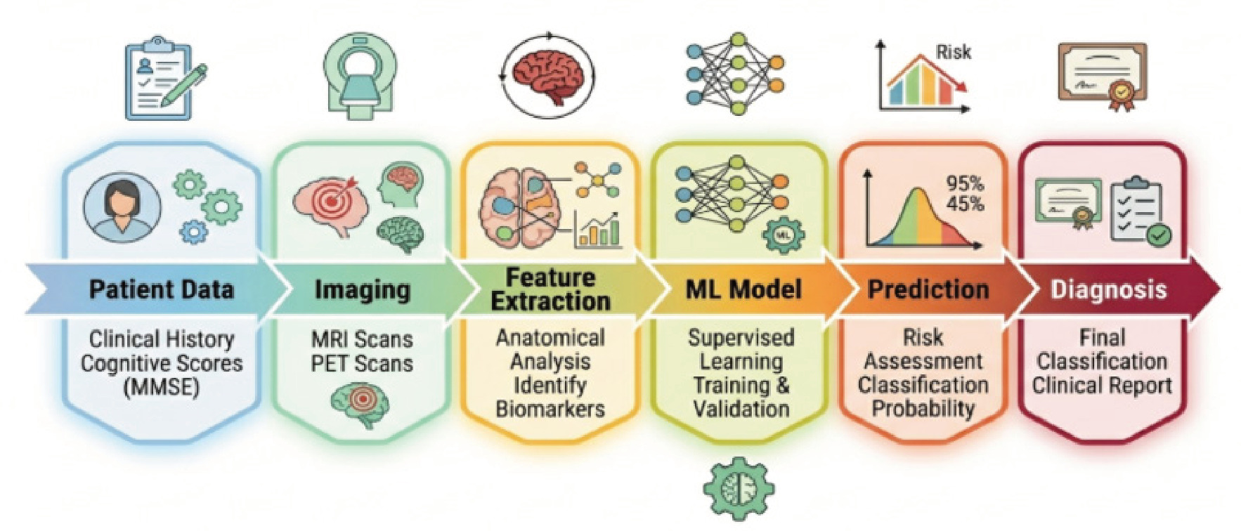
    AI technologies, biomarker-related advances and brain imaging technique, such as imaging biomarkers, have revolutionized our current diagnostic methods and treatment for AD patients. Many technologies have showed great promising, however, further changes for a faster, accurate and early detection for disease prediction, and individually personalized for the treatment in future research could be foreseen. In the recent study, there are significant efforts to establish the composite technology based on biologic information, clinical data and technology information system for precision care . Many promising technologies for shifting current care from symptom assessment towards predictive, preventative medicine will bring opportunities for patients and future study for AD with individualized, multi-modal marker integrated and explainable AI based, wearable healthcare technology. Personalized Medicine A leading frontier in Alzheimer's disease diagnosis and treatment is personalised medicine. Today, therapies for patients typically provide broad similarities to an assortment of patients irrespective of their genetics, stages of illness and how these individuals answer therapeutic responses. Nonetheless, Alzheimer's has numerous differences among sufferers from a bio-standard point of reference point -making it increasingly important for personalized medications. Personalised medicine strives to create healthcare measures for each individual's unique attributes including genotype, biomarkers patterns, family members health record and day-to-day lifestyles. Innovations made within Artificial Intelligence (AI) can help recognize distinctive markers of sickness to give each affected individual particular clinical and treatment options strategies. For example, an AI application could analyze specific knowledge collected via body biomarkers, cerebral spinal fluid markers, brain imaging information and genetic info to anticipate health progress and therapeutic replies, helping medical professionals determine viable alternatives and attain superior treatments effects. Personalized medication might even contribute in order to early precautionary programs . Using Artificial intelligence to discover people with the elevated chance before signs appear, personalized treatments (such as lifestyle improvements and neurological education) may help postpone condition development. Multi-Modal Biomarkers Future approaches to Alzheimer's disease diagnosis will involve combining different biomarker types instead of relying on a single biomarker . Although individual biomarkers may offer useful insights into certain aspects of the disease, none are adequate to encompass the whole range of Alzheimer's disease processes. By considering several biomarkers together, from cerebrospinal fluid or blood samples, genetic data or neuroimaging, the understanding of how the disease progresses and who is susceptible might improve substantially, contributing to early diagnostic decisions. Using data from structural MRI scans, PET imagery and additional biomarker information will also play an important role in developing the best diagnostic criteria . Explainable Artificial Intelligence In diagnostic AI systems with promising results, a primary challenge to clinical translation is transparency of deep learning models (many of which operate as black-boxes wherein the logic leading to the diagnosis is not easily explainable). Explainable Artificial Intelligence (XAI) refers to a set of research techniques that aim to make AI transparent and comprehensible. XAI enables understanding how the AI arrives at the diagnosis, while also revealing which input variables lead to a diagnosis/ risk assessment/etc. In the context of AD diagnosis, it allows for a deeper clinical understanding of why a certain diagnosis was reached for the given patient. As interpretability increases, clinician trust will improve and will ease assimilation into the clinical workflow of AD treatment. Moreover, XAI can aid in identifying bias of the algorithm and foster fairness in the clinical diagnosis process, also helping in clinical regulatory process validation. In the future, it would be desirable that the prediction performance is in balance with explainability in the models . Wearable Devices Another exciting area in future diagnosis and monitoring of Alzheimer's is the role of wearable healthcare technologies . The development of new sensor technologies and the advances in mobile health have led to ability to accumulate information about patients and behaviours in real world context using device that individuals use every day. Wearable tech like smartwatch, biosensors and portable monitoring systems which collect data on levels of physical activity, sleep characteristics, variability in heartbeat, movement patterns and even data relating to task and performance can be used to capture information which can indicate to subtle behavioural changes in the early stages of Alzheimer's disease. Algorithms AI powered analyses can use these generated data from wearable device and spot abnormal patterns that indicate potential decline in mental performance and therefore enhance early identification of a diagnosis . This technology can also be useful in the remote monitoring and telemedicine approach; where the number of visits to hospital can be reduced and improve accessibility for people who may be in isolated rural settings or older people. In conclusion the future diagnosis and treatment for Alzheimer's will rely on personalized approach, using different biomarkers to increase accuracy with transparent AI technologies as well as wearable technologies to achieve this. With the ongoing innovation and teamwork, the diagnosis of Alzheimer's disease can reach much more people, early detection will contribute to successful treatment of Alzheimer's disease, as shown in (Figure 4).

    CONCLUSION

    Currently, the most significant challenge to the medical health system is Alzheimer's disease, a type of neurodegenerative disorder of progressive nature and escalating global spread that causes tremendous Socioeconomic impact. Alzheimer's diseases' clinical diagnosis remains to be a crucial dilemma, as physical and pathological characteristics are usually initiated as early as many years before the manifestation of visible signs and symptoms of the illness. Current methods of diagnosis might not detect alterations associated with the Alzheimer's diseases at its early stages. It thus becomes mandatory to search for better advanced methods to diagnose and treat Alzheimer's disease in its early stages as early as possible to ensure efficient medical intervention and better patient management. Developments in Alzheimer's diseases Biomarker research and neuroimaging methods have remarkably increased our perception of the pathophysiology in Alzheimer's Diseases. Bio fluids obtained by Cerebrospinal Fluid (CSF), and blood analysis, genetic study and scanning images has become an essential indicator for the amyloid deposition and tau pathology. Furthermore, with growing interest in AI, analysis with huge set of data can be done at speed using machines through machine learning and deep learning approaches. Both the neuroimaging-based and the bio-fluid-based biomarkers for AD that use ML and DL has promised prospects and many studies have addressed the implementation and capabilities of those methods for the detection, classification, severity estimation of the AD. However, various technical constraints including over-fitting and under-fitting of model and insufficient data are yet needed to be solved. This review has broadly looked at the applications and capabilities of the neuroimaging and biomarkers in AD

    References

    Aghajanian, S., Mohammadifard, F., Mohammadi, I., Rajai Firouzabadi, S., Baradaran Bagheri, A. (2025). Longitudinal structural MRI-based deep learning and radiomics features for predicting Alzheimer's disease progression. Alzheimer's Research and Therapy, 17(1), 182.
    Ali, H. (2023). Artificial intelligence in multi-omics data integration: Advancing precision medicine, biomarker discovery and genomic-driven disease interventions. Int J Sci Res Arch, 8(1), 1012-30.
    Almughamisi, M., Amer, F., Alqahtani, N. K., Felemban, A. H., Jawharji, M. T. (2026). Differential effects of fresh and black garlic extracts on oxidative stress regulation and anti-inflammatory response properties in human HepG2 cells: An in vitro study. Frontiers in Pharmacology, 17, 1776975.
    Arya, A. D., Verma, S. S., Chakarabarti, P., Chakrabarti, T., Elngar, A. A. (2023). A systematic review on machine learning and deep learning techniques in the effective diagnosis of Alzheimer's disease. Brain Informatics, 10(1), 17.
    Ashton, N. J., Keshavan, A., Brum, W. S., Andreasson, U., Arslan, B. (2025). The Alzheimer's association Global Biomarker Standardization Consortium (GBSC) plasma phospho-tau round robin study. Alzheimer's and Dementia, 21(2), e14508.
    Bacon, E. J., He, D., Achi, N. B. A. D. A., Wang, L., Li, H. (2024). Neuroimage analysis using artificial intelligence approaches: a systematic review. Medical and Biological Engineering and Computing, 62(9), 2599-627.
    Beniczky, S., Schomer, D. (2020). Electroencephalography: basic biophysical and technological aspects important for clinical applications. Epileptic Disorders, 22(6), 697-715.
    Ben-Jaafar, A., Sinha, A., Nkrumah-Boateng, P. A., Roy, S., Sanker, V. (2026). Artificial intelligence-based biomarkers for the diagnosis and treatment of neurological conditions: a narrative review. Molecular Brain, 19(1), 26.
    Blennow, K. (2017). A review of fluid biomarkers for Alzheimer's disease: moving from CSF to blood. Neurology and therapy, 6(Suppl 1), 15-24.
    Chen, T., Shang, C., Su, P., Keravnou-Papailiou, E., Zhao, Y. (2021). A decision tree-initialised neuro-fuzzy approach for clinical decision support. Artificial Intelligence in Medicine, 111, 101986.
    Chen, X. Q., Mobley, W. C. (2019). Alzheimer disease pathogenesis: insights from molecular and cellular biology studies of oligomeric Aβ and tau species. Frontiers in Neuroscience, 13, 659.
    Chudzik, A., Śledzianowski, A., Przybyszewski, A. W. (2024). Machine learning and digital biomarkers can detect early stages of neurodegenerative diseases. Sensors, 24(5), 1572.
    Cummings, J., Aisen, P. S., DuBois, B., Frölich, L., Jack Jr, C. R. (2016). Drug development in Alzheimer's disease: the path to 2025. Alzheimer's research and therapy, 8(1), 39.
    Dara, O. A., Lopez-Guede, J. M., Raheem, H. I., Rahebi, J. (2023). Alzheimer's disease diagnosis using machine learning: a survey. Applied Sciences, 13(14), 8298.
    De Haan, W., Pijnenburg, Y. A., Strijers, R. L., van der Made, Y. (2009). Functional neural network analysis in frontotemporal dementia and Alzheimer's disease using EEG and graph theory. BMC neuroscience, 10(1), 101.
    De Leon, M. J., DeSanti, S., Zinkowski, R., Mehta, P. D. (2004). MRI and CSF studies in the early diagnosis of Alzheimer's disease. Journal of internal medicine, 256(3), 205-23.
    DeKosky, S. T., Marek, K. (2003). Looking backward to move forward: early detection of neurodegenerative disorders. Science, 302(5646), 830-34.
    DeTure, M. A., Dickson, D. W. (2019). The neuropathological diagnosis of Alzheimer's disease. Molecular neurodegeneration, 14(1), 32.
    Dhikav, V., Anand, K. (2011). Potential predictors of hippocampal atrophy in Alzheimer's disease. Drugs and aging, 28(1), 1-11.
    Dijkhuizen, R. M., van der Marel, K., Otte, W. M., Hoff, E. I., van der Zijden, J. P. (2012). Functional MRI and diffusion tensor imaging of brain reorganization after experimental stroke. Translational stroke research, 3(1), 36-43.
    Dorszewska, J., Prendecki, M., Oczkowska, A., Dezor, M., Kozubski, W. (2016). Molecular basis of familial and sporadic Alzheimer's disease. Current Alzheimer Research, 13(9), 952-63.
    Fabrizio, C., Termine, A., Caltagirone, C., Sancesario, G. (2021). Artificial intelligence for Alzheimer's disease: promise or challenge?. Diagnostics, 11(8), 1473.
    Fortea, J., Pegueroles, J., Alcolea, D., Belbin, O., Dols-Icardo, O. (2024). APOE4 homozygosity represents a distinct genetic form of Alzheimer's disease. Nature medicine, 30(5), 1284-91.
    Fox, N. C., Schott, J. M. (2004). Imaging cerebral atrophy: normal ageing to Alzheimer's disease. The Lancet, 363(9406), 392-94.
    Galvin, J. E., Price, J. L., Yan, Z., Morris, J. C., Sheline, Y. I. (2011). Resting bold fMRI differentiates dementia with Lewy bodies vs Alzheimer disease. Neurology, 76(21), 1797-803.
    Ghaffar Nia, N., Kaplanoglu, E., Nasab, A. (2023). Evaluation of artificial intelligence techniques in disease diagnosis and prediction. Discover Artificial Intelligence, 3(1), 5.
    Grueso, S., Viejo-Sobera, R. (2021). Machine learning methods for predicting progression from mild cognitive impairment to Alzheimer's disease dementia: a systematic review. Alzheimer's research and therapy, 13(1), 162.
    Hansson, O., Lehmann, S., Otto, M., Zetterberg, H., Lewczuk, P. (2019). Advantages and disadvantages of the use of the CSF Amyloid β (Aβ) 42/40 ratio in the diagnosis of Alzheimer's Disease. Alzheimer's research and therapy, 11(1), 34.
    Hur, J. Y. (2022). γ-Secretase in Alzheimer's disease. Experimental and molecular medicine, 54(4), 433-46.
    Hussain, D., Al-Masni, M. A., Aslam, M., Sadeghi-Niaraki, A., Hussain, J. (2024). Revolutionizing tumor detection and classification in multimodality imaging based on deep learning approaches: Methods, applications and limitations. Journal of X-Ray Science and Technology, 32(4), 857-911.
    Inamdar, A., Bugadannavar, P., Palled, M., Umarani, S., Salve, P. (2025). Biological determinants of blood-based biomarker levels in Alzheimer's disease: Role of nutrition, inflammation, and metabolic factors. Frontiers in Aging Neuroscience, 17, 1614962.
    Ji, F., Pasternak, O., Liu, S., Loke, Y. M., Choo, B. L. (2017). Distinct white matter microstructural abnormalities and extracellular water increases relate to cognitive impairment in Alzheimer's disease with and without cerebrovascular disease. Alzheimer's Research and Therapy, 9(1), 63.
    Johnson, G. V., Stoothoff, W. H. (2004). Tau phosphorylation in neuronal cell function and dysfunction. Journal of cell science, 117(24), 5721-29.
    Kardjadj, M. (2025). Advances in point-of-care infectious disease diagnostics: integration of technologies, validation, artificial intelligence, and regulatory oversight. Diagnostics, 15(22), 2845.
    Kung, H. F. (2012). The β-amyloid hypothesis in Alzheimer's disease: seeing is believing. ACS medicinal chemistry letters, 3(4), 265-67.
    Laforce Jr, R., Soucy, J. P., Sellami, L., Dallaire-Théroux, C., Brunet, F. (2018). Molecular imaging in dementia: past, present, and future. Alzheimer's and Dementia, 14(11), 1522-52.
    Lai, Y., Lin, C., Lin, X., Wu, L., Zhao, Y., Lin, F. (2022). Identification and immunological characterization of cuproptosis-related molecular clusters in Alzheimer's disease. Frontiers in aging neuroscience, 14, 932676.
    Laws, S. M., Hone, E., Gandy, S., Martins, R. N. (2003). Expanding the association between the APOE gene and the risk of Alzheimer's disease: possible roles for APOE promoter polymorphisms and alterations in APOE transcription. Journal of neurochemistry, 84(6), 1215-36.
    Lepeta, K., Lourenco, M. V., Schweitzer, B. C., Martino Adami, P. V., Banerjee, P. (2016). Synaptopathies: synaptic dysfunction in neurological disorders-A review from students to students. Journal of neurochemistry, 138(6), 785-805.
    Mace, N. L., Rabins, P. V. (2025). The 36-hr day: A family guide to caring for people with Alzheimer disease, other dementias, and memory loss (8th ed.).
    Maleki Balajoo, S., Rahmani, F., Khosrowabadi, R., Meng, C., Eickhoff, S. B. (2022). Decoupling of regional neural activity and inter-regional functional connectivity in Alzheimer's disease: a simultaneous PET/MR study. European journal of nuclear medicine and molecular imaging, 49(9), 3173-85.
    Mankhong, S., Kim, S., Lee, S., Kwak, H. B., Park, D. H., Joa, K. L., Kang, J. H. (2022). Development of Alzheimer's disease biomarkers: From CSF-to blood-based biomarkers. Biomedicines, 10(4), 850.
    Marey, A., Arjmand, P., Alerab, A. D. S., Eslami, M. J., Saad, A. M., Sanchez, N., Umair, M. (2024). Explainability, transparency and black box challenges of AI in radiology: impact on patient care in cardiovascular radiology. Egyptian Journal of Radiology and Nuclear Medicine, 55(1), 183.
    Márquez, F., Yassa, M. A. (2019). Neuroimaging biomarkers for Alzheimer's disease. Molecular neurodegeneration, 14(1), 21.
    Mattsson, N., Insel, P. S., Palmqvist, S., Portelius, E., Zetterberg, H. (2016). Cerebrospinal fluid tau, neurogranin, and neurofilament light in Alzheimer's disease. EMBO molecular medicine, 8(10), 1184-96.
    Mier, W., Mier, D. (2015). Advantages in functional imaging of the brain. Frontiers in human neuroscience, 9, 249.
    Mohammed, S., Malhotra, N. (2025). Ethical and regulatory challenges in machine learning-based healthcare systems: A review of implementation barriers and future directions. BenchCouncil Transactions on Benchmarks, Standards and Evaluations, 5(1), 100215.
    Mohsen, S. (2025). Alzheimer's disease detection using deep learning and machine learning: a review. Artificial Intelligence Review, 58(9), 262.
    Moloney, C. M., Lowe, V. J., Murray, M. E. (2021). Visualization of neurofibrillary tangle maturity in Alzheimer's disease: a clinicopathologic perspective for biomarker research. Alzheimer's and dementia, 17(9), 1554-74.
    Mosconi, L., Pupi, A., De Leon, M. J. (2008). Brain glucose hypometabolism and oxidative stress in preclinical Alzheimer's disease. Annals of the new York Academy of Sciences, 1147(1), 180-95.
    Nguyen, D., Nguyen, H., Ong, H., Le, H., Ha, H. (2022). Ensemble learning using traditional machine learning and deep neural network for diagnosis of Alzheimer's disease. IBRO Neuroscience Reports, 13, 255-63.
    Pagallo, U., O'Sullivan, S., Nevejans, N., Holzinger, A., Friebe, M. (2024). The underuse of AI in the health sector: opportunity costs, success stories, risks and recommendations. Health and technology, 14(1), 1-14.
    Patel, U. K., Anwar, A., Saleem, S., Malik, P., Rasul, B., Patel, K., Arumaithurai, K. (2021). Artificial intelligence as an emerging technology in the current care of neurological disorders. Journal of neurology, 268(5), 1623-42.
    Patharkar, A., Cai, F., Al-Hindawi, F., Wu, T. (2024). Predictive modeling of biomedical temporal data in healthcare applications: review and future directions. Frontiers in physiology, 15, 1386760.
    Phelps, M. E. (2000). Positron emission tomography provides molecular imaging of biological processes. Proceedings of the National Academy of Sciences, 97(16), 9226-33.
    Pritam, P., Deka, R., Bhardwaj, A., Srivastava, R., Kumar, D. (2022). Antioxidants in Alzheimer's disease: Current therapeutic significance and future prospects. Biology, 11(2), 212.
    Puntmann, V. O. (2009). How-to guide on biomarkers: biomarker definitions, validation and applications with examples from cardiovascular disease. Postgraduate medical journal, 85(1008), 538-45.
    Qiu, C., Kivipelto, M., Von Strauss, E. (2009). Epidemiology of Alzheimer's disease: occurrence, determinants, and strategies toward intervention. Dialogues in clinical neuroscience, 11(2), 111-28.
    Rademakers, R., Cruts, M., Van Broeckhoven, C. (2003). Genetics of early-onset Alzheimer dementia. The Scientific World Journal, 3(1), 497-519.
    Sagkriotis, A. (2025). Real-world evidence and the future of personalized medicine: a global perspective on data, ethics, and equity. Frontiers in Health Services, 5, 1682159.
    Shin, H. C., Orton, M. R., Collins, D. J., Doran, S. J., Leach, M. O. (2012). Stacked autoencoders for unsupervised feature learning and multiple organ detection in a pilot study using 4D patient data. IEEE transactions on pattern analysis and machine intelligence, 35(8), 1930-43.
    Silva-Spínola, A., Baldeiras, I., Arrais, J. P., Santana, I. (2022). The road to personalized medicine in Alzheimer's disease: The use of artificial intelligence. Biomedicines, 10(2), 315.
    Sorour, S. E., El-Mageed, A. A. A., Albarrak, K. M., Alnaim, A. K. (2024). Classification of Alzheimer's disease using MRI data based on Deep Learning Techniques. Journal of King Saud University Computer and Information Sciences, 36(2), 101940.
    Spasov, S., Passamonti, L., Duggento, A., Lio, P. (2019). A parameter-efficient deep learning approach to predict conversion from mild cognitive impairment to Alzheimer's disease. Neuroimage, 189, 276-87.
    Spies, P., Slats, D., Sjogren, J. M. C., Kremer, B. P. H., Verhey, F. R. J. (2010). The cerebrospinal fluid amyloid β42/40 ratio in the differentiation of Alzheimer's disease from non-Alzheimer's dementia. Current Alzheimer Research, 7(5), 470-76.
    Stancu, I. C., Vasconcelos, B., Terwel, D., Dewachter, I. (2014). Models of β-amyloid induced Tau-pathology: the long and "folded" road to understand the mechanism. Molecular neurodegeneration, 9(1), 51.
    Toumaj, S., Heidari, A., Shahhosseini, R., Jafari Navimipour, N. (2024). Applications of deep learning in Alzheimer's disease: a systematic literature review of current trends, methodologies, challenges, innovations, and future directions. Artificial Intelligence Review, 58(2), 44.
    Tsitsopoulos, P. P., Marklund, N. (2013). Amyloid-β peptides and tau protein as biomarkers in cerebrospinal and interstitial fluid following traumatic brain injury: a review of experimental and clinical studies. Frontiers in neurology, 4, 79.
    van Harten, A. C., Wiste, H. J., Weigand, S. D., Mielke, M. M., Kremers, W. K. (2022). Detection of Alzheimer's disease amyloid beta 1-42, p-tau, and t-tau assays. Alzheimer's and Dementia, 18(4), 635-44.
    Vemuri, P., Jack Jr, C. R. (2010). Role of structural MRI in Alzheimer's disease. Alzheimer's research and therapy, 2(4), 23.
    Viswan, V., Shaffi, N., Mahmud, M., Subramanian, K., Hajamohideen, F. (2024). Explainable artificial intelligence in Alzheimer's disease classification: A systematic review. Cognitive Computation, 16(1), 1-44.
    Vrahatis, A. G., Skolariki, K., Krokidis, M. G., Lazaros, K., Exarchos, T. P., Vlamos, P. (2023). Revolutionizing the early detection of Alzheimer's disease through non-invasive biomarkers: the role of artificial intelligence and deep learning. Sensors, 23(9), 4184.
    Wahul, R. M., Ambadekar, S., Dhanvijay, D. M., Dhanvijay, M. M., Dudhedia, M. A. (2025). Multimodal approaches and AI-driven innovations in dementia diagnosis: a systematic review. Discover Artificial Intelligence, 5(1), 96.
    Wang, C., Zong, S., Cui, X., Wang, X., Wu, S. (2023). The effects of microglia-associated neuroinflammation on Alzheimer's disease. Frontiers in immunology, 14, 1117172.
    Weissler, E. H., Naumann, T., Andersson, T., Ranganath, R., Elemento, O. (2021). The role of machine learning in clinical research: transforming the future of evidence generation. Trials, 22(1), 537.
    Yang, B., Yin, K., Lacasse, S., Liu, Z. (2019). Time series analysis and long short-term memory neural network to predict landslide displacement. Landslides, 16(4), 677-94.
    Yang, P., Bi, G., Qi, J., Wang, X., Yang, Y., Xu, L. (2025). Multimodal wearable intelligence for dementia care in healthcare 4.0: A survey. Information Systems Frontiers, 27(1), 197-214.
    Yuan, A., Nixon, R. A. (2021). Neurofilament proteins as biomarkers to monitor neurological diseases and the efficacy of therapies. Frontiers in neuroscience, 15, 689938.

    Acknowledgments

    The authors sincerely acknowledge Medi-Caps University, Indore, Madhya Pradesh, India, for providing the academic environment and library facilities that supported the preparation of this review manuscript.

    Author Contributions

    Shashank Sharma: Conceptualization, literature search, data curation, manuscript writing (original draft), visualization, and final manuscript preparation; Rupali Sontakke: Supervision, critical review, editing, validation, and intellectual input; Sanjay Jain: Conceptual guidance, manuscript review, supervision, and final approval of the manuscript. All authors have read and approved the final version of the manuscript.

    Conflict of Interest

    The authors declare that there is no conflict of interest.

    Related Articles